How advanced cardiac imaging can detect heart disease before symptoms appear
Heart disease may seem like something to worry about later in life. But changes inside your arteries can begin decades before chest pain or noticeable symptoms appear.
That’s one reason advanced cardiac imaging has become an important tool for evaluating coronary artery disease. Technologies such as coronary CT angiography, AI-assisted plaque analysis, and CT-derived fractional flow reserve (CT-FFR) show doctors a detailed look at plaque inside the heart’s arteries. In some cases, these tools can also show whether plaque is interfering with blood flow.
Heart disease can start earlier than you think
Coronary heart disease is a leading cause of death worldwide. The underlying disease process, called atherosclerosis, happens when fatty, inflammatory deposits known as plaque start building up within the walls of the coronary arteries.
“The process starts early and progresses silently over decades before physical symptoms or acute events happen,” says Nebraska Medicine cardiologist Kaveh Rezaei Bookani, MD.
This means some people may not feel sick while plaque develops in their arteries.
Identifying and addressing heart disease risk factors earlier in adulthood can help slow plaque formation and progression before coronary artery disease reaches a more advanced stage.
What are the early signs of coronary artery disease?
One of the challenges of coronary artery disease is that there may not be obvious early symptoms.
Symptoms often don’t develop until an artery has become significantly narrowed or if plaque ruptures, possibly causing a heart attack.
Some warning signs include:
- Chest pain, pressure, or discomfort during physical activity
- Shortness of breath during exertion
- Persistent or unusual fatigue
- A noticeable decline in your ability to exercise or do normal activities
These symptoms can be mistaken for stress, aging, or being out of shape. New, unexplained, or worsening symptoms should be discussed with your health care provider.
Call 911 for symptoms that could indicate a heart attack, including severe or persistent chest pain or pressure, especially when accompanied by shortness of breath, sweating, nausea, lightheadedness, or pain spreading to the arm, back, neck, or jaw.
Know your risk factors
High cholesterol, high blood pressure, diabetes, smoking, obesity, and physical inactivity are well-known risk factors for coronary artery disease. But there are other factors people may not associate with heart health.
Chronic inflammation, autoimmune conditions, sleep apnea, prolonged mental stress, and poor sleep quality can contribute to cardiovascular risk. These factors aren’t always reflected by a single blood pressure reading or cholesterol test.
Genetics make a difference too, but having a family history of heart disease doesn’t mean it’s predetermined.
While you can’t change your genes, other risk factors can be modified. Eating a nutrient-rich diet, getting regular physical activity, avoiding tobacco, getting enough sleep and managing blood pressure, cholesterol, and metabolic health can all help protect your heart.
What is a coronary CT angiogram?
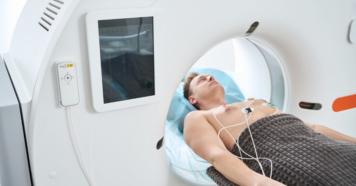
Also called coronary CT angiography, or CCTA, a coronary CT angiogram is a noninvasive imaging test that gives detailed images of the coronary arteries, the blood vessels that supply the heart muscle.
During the test, contrast material with iodine is administered through an IV. A CT scanner then captures images that can be reconstructed into detailed, three-dimensional views of the coronary arteries.
“These images allow physicians to look for plaque buildup and determine whether it is narrowing an artery,” says Dr. Bookani.
CCTA can also give information about plaque characteristics. For example, physicians can distinguish more heavily calcified plaque from noncalcified, or “soft,” plaque. Certain plaque features may be associated with a higher risk of rupture, which can lead to a heart attack.
How AI can help analyze plaque in the heart
A coronary CT angiogram produces a large amount of detailed information. AI-assisted plaque analysis can help physicians analyze those images further.
Machine learning algorithms can identify and measure coronary plaque seen on CT images. The technology can quantify how much plaque is present and help characterize different types of plaque, including calcified and noncalcified plaque.
“This gives physicians another way to evaluate a patient’s overall plaque burden instead of focusing only on whether one artery has developed a severe blockage,” says Dr. Bookani.
Clinical validation studies have shown high agreement between advanced CT analysis and invasive assessments. These tools are used with a physician’s interpretation and the patient’s overall clinical picture, not as a replacement for either.
Why finding nonobstructive plaque matters
A common misconception is that heart disease is important only once an artery is severely blocked.
Standard stress testing is especially useful for identifying coronary disease that’s progressed enough to reduce blood flow. But plaque can exist before it causes a severe blockage.
Advanced coronary CT imaging can identify plaque even when the narrowing is more mild.
“AI plaque analysis can then help quantify the total amount of plaque present,” says Dr. Bookani. “The additional information may help physicians identify patients with atherosclerosis earlier and determine whether preventive treatment or lifestyle changes should be intensified.”
In other words, the question isn’t always simply, “Is there a blockage?” It can also be, “How much coronary artery disease is already there?”
Who should consider coronary CT imaging?
Coronary CT angiography isn’t a routine screening test for every healthy young adult.
It is commonly used to evaluate people with symptoms that could be related to coronary artery disease. In certain circumstances, it can also be considered for people without symptoms who have a particularly high cardiovascular risk.
Whether CCTA is appropriate depends on your age, symptoms, medical history, risk factors, and other testing.
If you’re concerned about your heart disease risk, the first step is talking with your primary care provider or a cardiologist. They can assess your individual risk and determine whether additional testing or cardiac imaging makes sense.
Don’t wait for symptoms to start
The most important takeaway for younger adults isn’t that everyone needs advanced heart imaging. It’s that preventing heart disease starts long before most people think it does.
And when symptoms or risk factors need a closer look, advances in cardiac imaging can provide physicians with a much more detailed view of what’s happening inside the coronary arteries.
“We can identify plaque buildup, determine how much there is, and better understand whether it’s affecting blood flow,” says Dr. Bookani. “That information allows us to personalize treatment and potentially intervene before disease reaches a more advanced stage.”
Talk with your health care provider about your cardiovascular risk and whether evaluation by a Nebraska Medicine heart specialist is right for you. Schedule an appointment online or call 800.922.0000.