How does an ultrasound predict your due date and other answers to your ultrasound questions
Ultrasounds in pregnancy help providers monitor your baby’s health. They can also help predict due dates, reveal your baby’s biological sex, check for complications and more.
Read on for answers to your top ultrasound questions.
How does an ultrasound work?
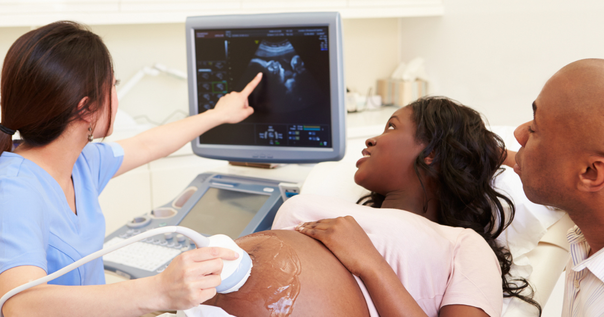
An ultrasound is also known as a sonogram. During an ultrasound, a sonographer uses a transducer to send and detect high-frequency sound waves.
In pregnancy, these sound waves bounce off a patient’s reproductive organs and the baby. The waves are then transformed into images that your provider sees on screen.
What happens during an ultrasound?
According to lead ultrasound technologist Brianna Heneger, BS, RDMS, RT, “The patient lays on their back, then we tuck them in with some towels to keep the gel we use from getting on clothes. We use ultrasound gel to keep contact with the probe and the patient’s skin. The sound waves from the probe don’t move through the air, so we need to stay in contact, or we’ll have dropout in our image. And then we use the probe to get the images you see on the screen.”
In the Olson Center for Women’s Health, you will typically meet a doctor for a brief discussion about the findings, which could include an estimated due date, the baby’s biological sex or an estimate of your baby’s weight.
What are the different types of ultrasounds?
In pregnancy, two types of ultrasounds are used:
- Transabdominal, during which the probe is moved over the belly
- Transvaginal, during which the probe is inserted vaginally
“We use a transvaginal ultrasound sometimes in the first trimester as things are smaller or if we can’t see through the abdominal wall,” Heneger says. “We occasionally use transvaginal later in pregnancy if we need to look at the cervical length with better detail or at the placenta’s location.”
When are ultrasounds usually done during pregnancy?
The number of ultrasounds you receive will vary, but common ultrasounds in pregnancy include:
- First-trimester screening, which is usually done around 11 to 13 weeks to:
- Document the baby’s heartbeat
- Measure the baby crown-to-rump to ensure an appropriate due date
- Screen for abnormalities if a patient desires or if there is a risk based on the mother’s or family history
- Anatomy scan, which is usually done around 18 to 20 weeks to:
- Check baby head-to-toe for anomalies
- Screen for some genetic conditions
- Measure amniotic fluid levels
“We’re trying to get patients in early in their pregnancies, but right now, during what is considered a ‘normal’ pregnancy, you’re really only guaranteed the 20-week anatomy scan,” Heneger says.
You may receive additional ultrasounds if:
- You experience bleeding, cramping or other concerns
- You are age 35 or older
- Your BMI is 35 or higher
- You have chronic hypertension or diabetes
- You have twins
When can an ultrasound show the sex of the baby?
Genital characteristics can often be seen by the 20-week anatomy scan, depending on the baby’s position. Be sure to tell your sonographer if you don’t want to know!
How does an ultrasound predict your due date?
During the first trimester, or before 14 weeks, an ultrasound can help predict the due date based on the crown-rump measurement of the baby. This is a straight-line measurement from the baby’s head to their bottom.
Typically, the last menstrual period is used to predict the due date. If there is bleeding early in pregnancy or irregular periods , an ultrasound may be the best way to determine the due date.
“If you're someone who's tracking your period, and you have regular periods every 28 days, it's a great way to date your pregnancy,” Heneger says. “If you have irregular periods, or you’re taking a shot in the dark on when it might have been, you may need an early ultrasound to get that due date.”
While later ultrasound scans can be used to predict due dates, they are less accurate than a first-trimester ultrasound with a crown-to-rump measurement.
Do I need to drink water before an ultrasound?
Drinking water and plenty of it before your ultrasound appointment can help your sonographer better visualize things, especially early in the pregnancy. The bladder is used as a sound “window” in some parts of the examination. After initial imaging, you may be asked to empty your bladder if it is overly filled or causing discomfort.
Are home ultrasounds or ultrasounds done at nonmedical facilities recommended?
You may have seen ultrasound kits for sale online or in stores that offer 3D or 4D ultrasounds.
As tempting as these options might be, maternal-fetal medicine specialist Teresa Berg, MD, cautions, “Recreational ultrasound is not recommended by the American College of Obstetricians and Gynecologists or the American Institute of Ultrasound in Medicine.”